Skip to content

Research Projects

How tissue mechanics, cell shape and cell fate connect

A circle of three ideas, each linked both ways to the next: tissue mechanics and cell shape affect each other, cell shape and cell fate affect each other, cell fate and tissue mechanics affect each other. In the middle, the lab’s model systems: outlines of a frog, Xenopus, and of a person. Each arc, and the person, stands for one of the lab’s projects, listed after the circle.

  1. Forces, fate and form: How cell and tissue mechanics coordinate both cell fate and tissue morphogenesis
  2. When forces decide fate: Understanding how mechanical forces regulate the timing of cell fate acquisition
  3. Shape meets genes: Integrating scRNAseq data with morphological parameters to generate a multimodal model of cell fate acquisition
  4. Organoid models of disease: Establishing new models of human respiratory pathologies using 3D organoids

Xenopus

Our model organism

The embryonic epidermis of Xenopus is a mucociliary epithelium that shares key cellular and functional features with the human airway epithelium, including multiciliated and mucus-secreting cells. Its external development and optical accessibility make it a powerful model for investigating epithelial morphogenesis, cell fate specification, and tissue mechanics through live imaging and controlled mechanical perturbations. Building on our long-standing expertise with Xenopus, we use this model system to uncover the fundamental principles coordinating cell identity, tissue organization, and mechanical forces during development.

How cell and tissue mechanics coordinate both cell fate and tissue morphogenesis

We want to understand the interplay between mechanical forces exerted by progenitor cells and generated within tissue to coordinate development of specialized tissues.

About this project ↓

Integrating scRNAseq data with morphological parameters to generate a multimodal model of cell fate acquisition

We use computational methods to integrate morphological data describing cell shape changes and cell movement dynamics with scRNAseq data across the development of mucociliary epithelium.

About this project ↓

Understanding how mechanical forces regulate the timing of cell fate acquisition

We want to determine when and how externally applied forces (stretch or compression) impact development of mucociliary epithelium.

About this project ↓

Establishing new models of human respiratory pathologies using 3D organoids

We engineer three-dimensional human respiratory organoids from patient-derived tissue biopsies to investigate disease mechanisms and develop physiologically relevant models for respiratory disease research.

About this project ↓

How cell and tissue mechanics coordinate both cell fate and tissue morphogenesis

Multiciliated cell progenitor (actin, green) probes the mechanics of the surrounding tissue (actin, pink) with long actin-rich protrusions (filopodia).

We want to understand the interplay between mechanical forces exerted by progenitor cells and generated within tissue to coordinate development of specialized tissues.

While a tissue forms, its cells constantly push and pull on each other. Young cells that have not yet chosen what to become, called progenitors, add forces of their own as they move into place. We want to know how these forces work together to build a tissue that does its job.

In the skin of frog embryos, new ciliated cells squeeze up into the outer layer and feel how stiff it is with thin, finger-like protrusions. We image this process in living embryos and use techniques such as atomic force microscopy (AFM) to measure mechanical forces and analyze tissue mechanics.

Papers from this project

  • Multiciliated cells use filopodia to probe tissue mechanics during epithelial integration in vivo

    Guilherme Ventura, Aboutaleb Amiri, Raghavan Thiagarajan, Mari Tolonen, Amin Doostmohammadi, Jakub Sedzinski

    Nature Communications, 2022 Publication

  • PAK4: A vanguard of epithelial vertex remodeling

    Raghavan Thiagarajan, Jakub Sedzinski

    The Journal of Cell Biology, 2026 Publication

  • ForSys: Non-invasive stress inference from time-lapse microscopy

    Augusto Borges, Jerónimo R. Miranda-Rodríguez, Alberto S. Ceccarelli, Guilherme Ventura, Jakub Sedzinski, Hernán López-Schier, Osvaldo Chara

    iScience, 2025 Publication

  • StressNET: an adaptable deep-learning model for mechanical stress inference in tissues

    Nicolás Aldecoa Rodrigo, Augusto Borges, Jerónimo R. Miranda-Rodríguez, Guilherme Ventura, Jakub Sedzinski, Hernán López-Schier, Osvaldo Chara

    bioRxiv, 2026 Preprint

  • Protocol for inferring mechanical stresses in tissues using ForSys, an open-source Python tool

    Augusto Borges, Jerónimo R. Miranda-Rodríguez, Alberto S. Ceccarelli, Guilherme Ventura, Jakub Sedzinski, Hernán López-Schier, Osvaldo Chara

    STAR Protocols, 2026 Publication

  • Emerging concepts on the mechanical interplay between migrating cells and microenvironment in vivo

    Guilherme Ventura, Jakub Sedzinski

    Frontiers in Cell and Developmental Biology, 2022 Publication

  • Emergence of an Apical Epithelial Cell Surface In Vivo

    Jakub Sedzinski, Édouard Hannezo, Fan Tu, Maté Biro, John B. Wallingford

    Developmental Cell, 2016 Publication

  • RhoA regulates actin network dynamics during apical surface emergence in multiciliated epithelial cells

    Jakub Sedzinski, Édouard Hannezo, Fan Tu, Maté Biro, John B. Wallingford

    Journal of Cell Science, 2017 Publication
Understanding how mechanical forces regulate the timing of cell fate acquisition

Inferring mechanical forces during tissue stretching and deformation in the developing epithelium.

We want to determine when and how externally applied forces (stretch or compression) impact development of mucociliary epithelium.

During development, the mucociliary epithelium experiences dynamic mechanical forces as surrounding tissues grow and remodel. We investigate how stretching and compression influence cell fate decisions and whether their effects depend on developmental timing. By applying controlled mechanical perturbations at defined stages, we aim to identify critical windows of mechanosensitivity and uncover how physical forces regulate epithelial cell differentiation.

Integrating scRNAseq data with morphological parameters to generate a multimodal model of cell fate acquisition

Live imaging reveals how cells dynamically change their shape and movement during epithelial development.

We use computational methods to integrate morphological data describing cell shape changes and cell movement dynamics with scRNAseq data across the development of mucociliary epithelium.

Each new cell in a developing airway lining becomes one of a few types, such as cells that grow cilia and cells that make mucus. Two kinds of data describe how. Movies show how every cell changes shape and moves. Single-cell RNA sequencing (scRNAseq) reads which genes each cell is using.

By integrating live imaging with single-cell transcriptomics, we investigate how cells acquire their identities during epithelial development. Our research reveals that dynamic changes in cell shape can predict future cell fate, while distinct cell types emerge progressively from shared progenitor states rather than following rigid, predetermined lineages.

Papers from this project

  • Single-cell morphodynamics predict cell fate decisions during mucociliary epithelial differentiation

    Mari Tolonen, Ziwei Xu, Ozgur Beker, Varun Kapoor, Bianca Dumitrascu, Jakub Sedzinski

    Molecular Systems Biology 22(7), 1040–1069, 2026 Publication

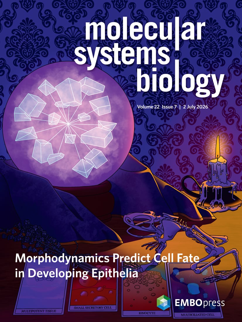
    On the cover of Molecular Systems Biology

    Cover of Molecular Systems Biology, July 2026: a crystal ball holding cell shapes above a frog skeleton and tarot cards naming the cell types
  • A single-cell, time-resolved profiling of Xenopus mucociliary epithelium reveals nonhierarchical model of development

    Julie Lee, Andreas Fønss Møller, Shinhyeok Chae, Alexandra Bussek, Tae Joo Park, Youni Kim, Hyun-Shik Lee, Tune H. Pers, Taejoon Kwon, Jakub Sedzinski, Kedar Nath Natarajan

    Science Advances, 2023 Publication

Establishing new models of human respiratory pathologies using 3D organoids

Human respiratory organoids grown from patient-derived tissue.

We engineer three-dimensional human respiratory organoids from patient-derived tissue biopsies to investigate disease mechanisms and develop physiologically relevant models for respiratory disease research.

We develop reproducible and scalable 3D human respiratory organoids from patient-derived tissues for disease modelling and high-throughput compound screening. By integrating quantitative measurements of organoid mechanical properties, we investigate how changes in tissue mechanics contribute to pathological epithelial remodeling in respiratory diseases, including asthma, COPD, and pulmonary fibrosis.

Papers from this project

  • Effects of n-3 Long-Chain Polyunsaturated Fatty Acid and Vitamin D Supplementation on Transcriptional Profiles of Human Lung Organoids

    Mina Ali, Martin Steen Mortensen, Ole Bæk, Nicklas Brustad, Tingting Wang, Liang Chen, Min Gyu Kim, Casper-Emil Tingskov Pedersen, Trevor D. Lawley, Athanasios Pasias, Jakub Sedzinski, Jakob Stokholm, Klaus Bønnelykke, Bo Chawes

    Metabolites, 2025 Publication